A.
UNDERSTANDING THE DEFINITION OF TRACHEA
Trachea is a pipe-shaped
tube organ that is part of the respiratory system with a length of 10-16 cm and
a diameter is about 20-25 mm. trachea lies before bronchi, After the larynx and
adjacent to the esophagus. The trachea is an organ that serves to distribute
air into the bronchi and alveoli , also simultaneously filter out dust or dirt
component in the air. Tracheal shape in living organisms can vary, but in
humans it is as described above.
B.
THE FUNCTIONS OF TRACHEA
1. Part of The Respiratory System
The trachea is
located after the breathing tube (larynx). Air that passes through trachea will
go to the bronchus, then to alveoli and then to the pulmonary. The dust or any
unsaved component in the air will be filtered by trakea. Trachea also can
maintain the air humidity as well as participate in regulating air temperature
because it has mucus (mucus) on the mucosa layer.
2. Participate on The Digestion Process
Most of the
tracheal wall are attached into the wall of the digestive organs, esophagus. So
indirectly trachea also have an influence on the digestive process in humans.
If there is something that block trachea then that is would be a problem too
for the esophagus. For example during the airway obstruction you will choke and
conduct the cough reflex so that the trachea and esophagus clear from foreign
objects.
3. Prevent Harmful Substances to Enter The
Lungs (To Protect The Respiratory Tract)
When there are
foreign objects that enter through the respiratory tract and to the trachea,
the object will be trapped and attached to the sticky mucus of trachea. Then
the object or the bacteria will be removed from the body in the form of a
liquid or viscous sputum (because it is mixed with trachea mucus).
The trachea is a
tube formed by 16-20 cm C-Shape cartilagous ring. This ring is not circular
because the ends do not converge due to attachment of the esophagus to the
tracheal wall. In addition, it is also useful that the trachea remains open and
can make a few changes in diameter when it is needed so that the air going in
and out smoothly. These rings are also tied together with fribrosa tissue. The
trachea is strong but also elastic. The trachea was composed by the ciliated
epithelium which have goblet cells, these goblet cells produce mucus (a thick
liquid) that protects the tracheal wall.In the section near to the lungs,
trachea structure forming two branches (left and right) which will connected
directly with the bronchi, alveoli and lung.
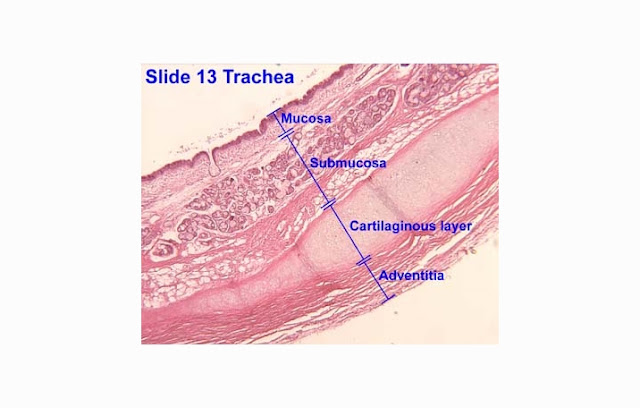
Tracheal wall
composed by three layers: (from inside out):
1. Mucous Layer (Inner Layer)
Mucosal layer of
the trachea composed by cylindrical ciliated epithelial cells with goblet
cells. This layer serves to produce extra mucus (viscous liquid) that protects
the tracheal wall and also protect the respiratory tract from foreign bodies.
2. Muscle Tissue and Cartilage (Middle
Layer)
Cartilage layer
is a layer of C-Shaped cartilage as we have explained previously. The opening
in the cartilage is located on the posterior (the meeting point of the trachea
with the esophagus). Around this cartilage layer, there are muscle tissue in
the form of smooth muscle, its function is to the control the movement of
breathing, coughing or choking reflex. In this layer there is also a structure
that connects one ring to another and keep both ends of the ring remains in an
optimal state.
3. Adventitia layer (Outer Layer)
The outermost
layer is composed by connective tissue. this layer also containing blood
vessels, nerves, and fat tissue.
 |
| TRACHEA LAYERS |
Several other
sources also mention that the tracheal wall has four layers, one more layer in
question is submucosal layer which located after Mucous Layer. Submucosal layer
is composed of connective tissue that looks apart from the epithelium of the
mucosal layer. In this layer can be found a lot of blood vessels and nerves.
This layer allows the movement of the tracheal mucous.


