A. UNDERSTANDING THE DEFINITION OF SMALL
INTESTINE
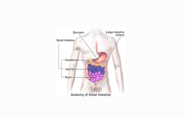
Small bowel (intestine) is one of the major digestive
part located after the stomach. Small intestine shape is narrow winding tube
that fill the lower abdomen cavity. The main function of the small intestine is
to perform chemical digestion and absorption of food. The small intestine has a
diameter of more than 2 cm long and about 6 meters in adults. The small
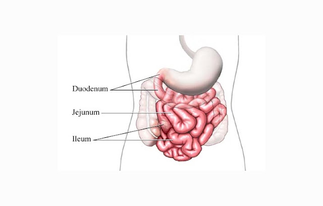
intestine is divided into three parts : the duodenum, jejunum, and Ileum. The
small intestine is often referred to as the longest digestive organs in the
human body.
B. THE FUNCTIONS OF SMALL INTESTINE
As we have explained above, the main function of the
small intestine is to absorb and digest food from the stomach. However, each
parts of the small intestine have their respective functions:
- The Duodenum, serves to break down the components of the stomach into smaller components that can be used by the body.
- The Jejunum, serves to do the digestion and absorption of various components, particularly, water, carbohydrates, protein, and vitamins, and other components that are lipophilic.
- The Ileum, serves for the absorption of salt, vitamin B and components of food that is not absorbed by the jejunum.
C. THE STRUCTURE OF SMALL INTESTINE
The small intestine has the same structure as other
digestive organs such as the stomach. The structure of the small intestine was
prepared by 4 walls (From outside to inside):
 |
| SMALL INTESTINE WALL |
1. Serous
Layer
The outermost layer is composed of blood vessels,
lymphatics and nerves. Serous layer of the small intestine is in the form of
connective tissue that is covered by visceral peritoneum. Serous layer has
small cavities where the discharge of serous fluid issued, this fluid serves as
a lubricant for muscle movement.
2. Muscle
Layer
Muscle layer of the small intestine is a layer of
smooth muscle that works involuntary. There are two types of muscle fibers, the
longitudinal muscle fibers (lengthwise) and circular muscle fibers (circular).
The combination of both types of muscle contraction will produce intestinal
peristalsis which serves to break down the food and took it to the next
digestive organ.
3. Submucosa
Layer
Layer that composed by connective tissue containing
blood vessels, lymphatics, nerves and mucous glands. The blood vessels in the
submucosal layer of the small intestine plays an important role in passing the
food and nutrition.
4. Mucosa
Layer
Mucous layer prepared by simple epithelial cells and
thin connective tissue. The mucosal lining had goblet cells that can produce
mucus. Mucus is a secretion from glands throughout the small intestine. The
production of this layer which influenced by the hormone secretin and
enterocrinin is often also called intestinal juice.
1. The
duodenum
The duodenum is part of the small intestine that is
located right after the stomach and before jejunum (Empty intestine). The
length of duodenum is approximately 25 cm (10 inch) - 38 cm (14.8 inch). The
duodenum is the shortest part of the small intestine compared to other
intestinal. This Part of start from the bulbo duodenale and ends by treitz
ligament. normal pH of the small intestine are 9. There are histological
structure called brunner glands that produce alkaline mucus to help the absorption
and neutralizing the pH of the food. Duodenum is retroperitoneal organ because
it is not entirely covered by the peritoneal membrane. Pancreatic and bile duct
are connected directly to the small intestine, pancreatic juice serves to break
down the food, while the bile juice is used to breakdown and digestion of fat. The
gastrointestinal products that enters the duodenum is called chyme. The
duodenum is used to treat, neutralize, break down and digest the chyme.
2. jejunum (Empty
intestine)
Jejunum is the middle part of the small intestine.
The length of the jejunum is from 1 - 2.5 meters. The word comes from modern
English language, the adjective "Jejune" which means hungry. this
understanding is derived from the Latin word "Jejunus" meaning empty.
Jejunum is hung and held by the mesentery (part of the lining of the
peritoneum), allows the jejunum to move during the process of digestion takes
place. Jejunum has a very large surface area, forming folds of intestines. On
the surface there is a finger-shaped protrusions called villi. This villi
serves for absorbing nutrients from food. The main function of the jejunum is
to break down the nutrition, absorption of lipophilic nutrients and absorb the
water. To differences between duodenum and jejunum can be seen with brunner
glands began to decrease when entering the jejunum and the growing number of
existing villi. But it is difficult to distinguish the jejunum and ileum
because they are macroscopically similar.
3. ileum
(Absorption Intestine)
Ileum is the last part of the small intestine. In the
human digestive system, ileum has a length of about 2-4 meters. ileum pH
ranging between 7 to 8 (neutral or slightly alkaline). In the ileum there is
also a protrusions-like structure called villi. Just as in the jejunum (Empty
intestine), villous serves to absorb nutrients such as sugars, amino acids,
fatty acids, glycerol, vitamins and minerals. Ileum serves to absorb vitamin B
(especially B12), bile salts, and the food is not absorbed in the jejunum.
E. ENZYMES ISSUED BY SMALL INTESTINE
- Enterokinase Enzymes, serves to convert trypsinogen to trypsin.
- Maltase Enzymes, to convert maltose into glucose and galactose.
- Sucrase Enzymes, serves to convert sucrose into glucose and fructose.
- Lipase intestine enzymes, serves to transform fat into fatty acids and glycerol.
- Erepsin / dipeptidase Enzymes, serves to convert peptones into amino acids.
- Disaccharidase enzymes, an enzyme that serves to convert disaccharides into monosaccharides.


