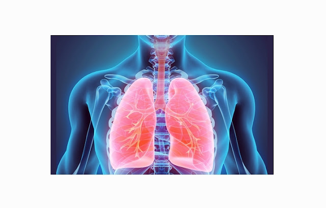
A. UNDESTANDING THE DEFINITION OF LUNGS
(PULMO)
Lungs is the main organ responsible for the process
of respiration (respiratory system in humans) and consists of two parts:
pulmonary dextra (right lung) and the left pulmonary (left lung). The lungs are
also associated with the circulatory system (circulation system) and excretion
(expenditure of wastes).
The lungs are a very important organ in the human
body, Its the place where the exchange of oxygen (O2) and carbon dioxide (CO2)
happen. In general, lungs, found in all mammals, including human.
B. PARTS OF LUNGS (PULMO)
The lungs are one of the vital organs in humans
located in the thoracic cavity (the chest). If the thoracic cavity is opened,
the volume of the lung can shrink up to 1/3 or less because it is very elastic
and are influenced by pressure changes. In children, lungs are pink and will be
accompanied by dark spots as you age due to inhalation of dust particles.
Anatomically, the lungs consists of the following
sections:
a. Apex (peak)
is called pulmonary apex, obtuse and stand up about 1 inch above the clavikula
(tullang collarbone) and is covered by the cervical pleura.
b. Three
surface, comprising:
- Costal surface: large / wide, soft, convex.
- Mediastinal surface: concave , contains hilum.
- Diaphragmatic surface: concave.
c. Three
restrictions, which consist of:
- anterior (front)
- inferior (bottom)
- posterior (back)
The lung is composed of two parts, the right lung
(pulmonary dextra) and the left lung (pulmonary sinistra). Both parts are
separated by a space which contains the heart and major blood vessels. This
space is called the mediastinum. The differences between these pulmonary
section are:
Right lung is shorter and wider than the left lung
due to the position of the diaphragm on the right side is higher than the left
side. Then, the grooves on the anterior margin of the left lung due to a heart
whose structure is shaped like a tongue (lingula). Thise grooves is called the
incisura cardiaca.
To be protected in the thoracic cavity, pulmo are
coated by a membrane called the pleura. The pleura is divided into two,:
- Visceral pleura, directly wraps the lung.
- Parietal pleura, membrane that lines the outside part of the chest cavity.
- Among both pleura, there are a cavity called the pleural cavity. The pleural cavity normally air vacuum and contains fluid, so that the lungs can expand and contract during breathing movements without friction because of the fluid (exudates) are useful to lubricate the surface of the pleura.
C. STRUCTURE OF LUNGS (PULMO)
In carrying out its functions, the lungs are
connected with several other organs :
- The trachea (windpipe), The air is inhaled from the nose and mouth will go throught trachea to enter the lung.Trachea starts from the larynx to the bronchus principalis dextra et sinitra.Diameter size of trachea is about ± 2.5 cm in adults and the size of a pencil diameter in infants.
- Bronchi, is a continuation of the trachea connecting the right lung and left lung. Bronchus is the air conduction channel taht directly related to the alveoli.
- Bronchioles, are branches of the bronchi, small tubes with a number of ± 30,000 pieces for one lung. This bronchioles will bring oxygen into the lungs.
- The alveoli, the ends of the bronchioles which number around 600 million in the adult human and is shaped like a small pouch. Alveoli walls are thin and consists of:
- Type I alveolar cells that form the structural basis;
- Type II alveolar cells that secrete surfactant serves to reduce surface tension between air and water.
- Immune cells called macrophages are also present in the alveoli to destroy pathogens and foreign trash. Alveolar wall has pores called Kohn pores, allowing air flow from the one alveoli into the other. Each of the alveoli is also surrounded by a network of capillaries that carry blood to the alveoli for the oxygenation process. In this aveoli, oxygen diffuses into carbon dioxide taken from the blood.
 |
| LUNGS STRUCTURE |
D. HOW DOES LUNGS (PULMO) WORKS
Air moves in and out of the lungs because the
difference of atmospheric pressure contained in the alveoli due to the
mechanical work of the muscles. During the process of inspiration, an increase
in volume is occured due to reduction of thoracic retraction of the diaphragm
and ribs. This occurs from contraction of several muscles : m.
sternocleidomastoid which serves to lift the sternum up, scalenus and lift intercostalis
external muscles.
During quiet breathing, expiration is passive because
the elasticity of the chest wall and the lung. At the time of the external
intercostal muscle relaxation, chest wall will go down and the arch of the
diaphragm will move up into the thoracic cavity, which causes the reduction of
thoracic cavity volume. This volume reduction intrapleura cause to increased
pressure and intrapulmonary pressure. This situation causes, the pressure
difference between the air duct and the atmosphere to be reversed, so that the
air flows out of the lungs until the air and atmospheric pressure are in
equation at the end of the expiration process.
The second phase of the breathing process includes
the process of diffusion of gases across the alveolar capillary membrane
(thickness of less than 0.5 m). The driving force for this transfer is the
partial pressure difference between the blood and the gas. The partial pressure
of oxygen in the atmosphere is about 149 mmHg. At the time we inhale oxygen and
reached the alveoli then this partial pressure will decrease to 103 mmHg. This
partial pressure drop occurs based on the fact that the inspired air is mixed
with air and water vapor in the room. The difference in pressure of carbon
dioxide in the blood with a much lower alveoli causing carbon dioxide diffuses
into the alveoli and later released into the atmosphere.
In a normal resting state, the diffusion and the
balance of oxygen in the blood capillaries of the lungs and alveoli lasts
approximately 0.25 seconds of total contact time for 0.75 seconds. It means
that normal lung have enough diffusions spare time.
E. PULMONARY FUNCTION - PULMONARY
Lung has a very important role. The main function of
the lung is for gas exchange between carbon dioxide in the blood with oxygen
from the atmosphere. The purpose of this is to provide oxygen that needed by
tissue and remove carbon dioxide. Air enters the lungs through a narrows pipe
system (bronchi and bronchioles) and trachea. The pipe ends in the lung
(alveoli) in the air pockets where oxygen and carbon dioxide removed. Based on these
functions, respiration process can be divided into four basic mechanisms,
namely:
- Ventilation, ie a process of entrance and exit of air between the atmosphere and the alveoli of the lungs;
- Diffusion, a process the influx of oxygen and remove carbon dioxide between the alveoli and blood;
- Transportation, which is a process to transport oxygen and carbon dioxide in the blood and body fluids to and from body tissue cells;
- Ventilation Regulation.
F. FACTORS
AFFECTING THE LUNG FUNCTION
Here are the factors that can affect the lungs :
1. Age
Maximum muscle strength is at the age of 20-40 years
and be reduced by 20% after the age of 40 years. During the aging process, the
elasticity of the alveoli is decreasing, the bronchial glands is thickening,
and decreased lung capacity.
2. Gender
Ventilation function in males is 20-25% higher than
women, because the size of the male anatomy lung are greater than women. In
addition, the activity of males is higher so that the recoil and lung
compliance are already trained.
3. Height and
weight
A person who has a high and large body has better ventilation
function than the smallish short person.


