A. UNDERSTANDING THE DEFINITION OF TENDON
Tendons are connective tissues that connect bone to
muscle. Tendon serves to balance the bone and muscle when the movement process.
Tendon that attached to the bone and mobile called insersio (insertian =
inset), while the tendon attached to the bone but immobile called Origo (origin
= origin). The structure of tendon at the tip muscles is small, tough and hard.
B. THE FUNCTIONS OF TENDON
As we have explained previously, the main function of
the tendon is to control the movement mechanism (effective and efficient). When
the movement occur, the tendon will adapt to the changing position of the bones
and muscles so that the movement is perfect. Tendon function is associated with
the contraction (shortening) and relaxation (elongation) of muscle. When the
muscles are contract, the tendons transmit energy from the contraction to the
bones and joints, while the muscles are relax, tendons will also adjust its
state. Although it has a very strong structure, tendon can also be damaged. Most
often Injuries to the tendon due to excessive training or accident.
C. THE STRUCTURE OF TENDON
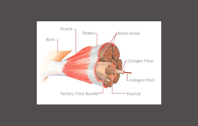
Please note the above image to make it easier to
understand our explanation
We will try to explain the structure of the tendon
form the smallest constituent components.
The smallest structure of tendon is collagen fibrils.
These fibrils are solid, strong and flexible. The nature of the collagen fibers
would make it resistant to traction and encouragement between the bones and
muscles. The basic constituent molecules of collagen fibrils is from some tropokolagen united to form microfibrils and microfibrils of
collagen are combined together to form subfibril
collagen, and then combined to form collagen
fibrils.
 |
| TENDON STRUCTURE |
The combination of collagen fibrils will merge and
form a protective layer in the form of strands called collagen fibers.
Then the combination of collagen fibers that are
coated by a endotendon layer will form Primary Collagen Fiber (Sub-fasicle).
The primary will be joining to each other and coated
by endotendon layer (layer that serves to protect and stabilize the tendon) to
form Secondary collagen fiber bundles (fasicle).
A combination of several secondary collagen fibers that
coated by endotendon layer (layer that serves to protect and stabilize the
tendon) formed Tertiary Collagen Fiber Bundles.
Now a collection of Tertiary Collagen Fiber Bundles
that coated by epitendon layer (the outer layer of the tendon) will form the complete
Tendon.


