Bone : Defintion, Structure, Functions, Types

A. UNDERSTANDING THE DEFINITION OF BONE
Bone is strong and resilient organs that give shape to the body. Boneless body will not be able to stand upright. Bone also serves in the motion system. In everyday life people are always doing the motion (movement). System of motion in humans is supported the two main components : bone and muscle. Muscle is called active locomotor and bone is called passive locomotor. The bone is a passive tool movement because they can not move themselves, but must be driven by muscle.
Definition, Structure and Functions of Bones
BONES
B. THE FUNCTIONS OF BONE
Eac Bone has a different shape. So the bone will function according to their shape. Here are some of the functions of human bones.
  • Supporting and giving body shape
  • Protector of the vital organ of the body such as the brain, heart, lungs etc.
  • Composer skeleton
  • Where the muscles attach
  • Points formation of blood cells
  • Storage of minerals (potassium and phosphorus)

C. TYPES OF BONE
Based on its constituent tissue, bone can be divided into two kinds : cartilage (soft bones) and hard bones (osteon).
1. The cartilage
When you hold the ear or nose tip, you can feel supple area. Why is that? Concha and the nasal tip feels supple because it was formed by cartilage.
Baby in the womb is almost entirely composed of cartilage. Cartilage will develop into hard bone, although not entirely. So that in adults, the cartilage is only found in certain places, such as earlobes, tip of the nose, larynx, trachea, bone joint surface, between the ribs and sternum, and between the segments of the spine.
Cartilage is formed by cartilage cells (chondrocytes) and matrix (base material). Cartilage can be divided into three kindsv : hyaline cartilage, elastic cartilage and fibrous cartilage.
  • Hyaline cartilage, is smooth, transparent, and has a homogeneous matrix. Hyaline cartilage can be found on the surface of the joints and the tracheal wall.
  • Elastic cartilage, is flexible and elastic, matrix containing fibers branching. Elastic cartilage found on the tip of the nose and earlobes.
  • Fibrous cartilage, is less flexible and contains many fibers of collagen matrix. Fibrous cartilage can be found between vertebrae segments and cartilage in the knee (tendons and ligaments).

2. Bone (osteon)
In contrast to the cartilage, the hard bone is composed by bone cells (osteocytes). Bone cells secrete matrix containing calcium and phosphorus so that bones become hard and inflexible. Bone matrix is ​​not solid and has hollow-empty cavity to form spongy bone. Most of the hard bone will draw up the framework of the body.

D. SHAPES OF THE BONE
Based on the shape, bone can be divided into four types : short bones, long bone, flat bones and irregular bones.
1. Long Bones
It has a shape like a hollow tube. Long Bones can be found in the femur, fibula, ulna, tibia, and lever.

2. Short bones
It has a shape like a cube. This bone is only found at the base of the foot, armpit, and the joints of the spine.

3. Flat Bone
As the name suggests, this form of the bones is flat. Flat bone serves as constituent or protector of a cavity in our body. Examples of flat bones are ribs, scapula, and skull.

4. Irregular Bones
Bones that have irregular shapes. Irregular bones can be found in the bones of the face and spine.
Shapes Of The Bone
BONE SHAPE
E. STRUCTURE OF THE BONE
Long bones such as the femur consists of two different structures : compact bone (cortical) and spongy bone (cancellous or trabecular). Compact bone form a solid cylinder on the central axis of the bone surrounding the bone marrow cavity. Accounted for 80% of bone mass in the human body is Compact Bones. Spongy bone located at the ends of long bones, accounted for about 20% of total bone mass and has a structure like a beehive.

Bone itself is made up of 10-20% water, about 60-70% mineral and the rest is collagen (Collagen is a major fibrous protein in the body), but bone also contains small amounts of other substances such as proteins and inorganic salts. The composition of the mineral component of bone form as hydroxyapatite (HA) which is a combination of calcium and phosphate, with the chemical formula CA10 (PO 4) 6 (OH) 2.

At the ends of the bones, there are an extension pipe function to connect on bone with another bone. The wide end of the bone which is composed of spongy bone called epifise. Mid section which extends between that wide end called diafise, its a compact bone containing a cavity called the bone marrow cavity. Between Diafise and epifise is an area called epifise chakras. Epifise chakra area is growing from time to time as long as someone is still growth.

F. FORMATION OF BONE
In the stage of embryonic development, our body is still a skeleton of cartilage. Cartilage is formed by the cells of the mesenchyme. In cartilage will be filled by osteoblasts. Osteoblast is A bone-forming cells. Osteoblasts will fill the surrounding tissue and form osteocytes (bone cells) .Bone cells is form concentric shapes (from the inside to the outside), each bone cells will surround the blood vessels and nerve fibers, create a system called Havers systems. Bone also has osteoclasts, These cells function is to reabsorb bone cells that have been damaged and destroyed. The existence of these osteoclast cell activity can causes bones to be hollow. This cavity will eventually be filled by the bone marrow.
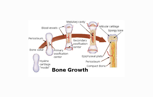
Bone Formation
FORMATION OF THE BONE
The process of bone formation is called ossification. The process is divided into two : intramembranosa ossification and intracartilagenosa ossification. Intramembranisa ossification also known as direct (primary ossification). This process occurs in flat bones such as the skull. Reinforcement is only done once and will not be repeated forever. And the example of intercartilagenosa is the formation of long bone that will increase bone length.
Lungs (Pulmo) : Definition, Structure and Functions

A. UNDESTANDING THE DEFINITION OF LUNGS (PULMO)
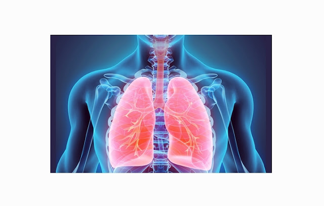
Lungs is the main organ responsible for the process of respiration (respiratory system in humans) and consists of two parts: pulmonary dextra (right lung) and the left pulmonary (left lung). The lungs are also associated with the circulatory system (circulation system) and excretion (expenditure of wastes).
The lungs are a very important organ in the human body, Its the place where the exchange of oxygen (O2) and carbon dioxide (CO2) happen. In general, lungs, found in all mammals, including human.
Definition, Structure and Function of Lungs (Pulmo)
LUNGS
B. PARTS OF LUNGS (PULMO)
The lungs are one of the vital organs in humans located in the thoracic cavity (the chest). If the thoracic cavity is opened, the volume of the lung can shrink up to 1/3 or less because it is very elastic and are influenced by pressure changes. In children, lungs are pink and will be accompanied by dark spots as you age due to inhalation of dust particles.
Anatomically, the lungs consists of the following sections:
a. Apex (peak) is called pulmonary apex, obtuse and stand up about 1 inch above the clavikula (tullang collarbone) and is covered by the cervical pleura.

b. Three surface, comprising:
  • Costal surface: large / wide, soft, convex.
  • Mediastinal surface: concave , contains hilum.
  • Diaphragmatic surface: concave.

c. Three restrictions, which consist of:
  • anterior (front)
  • inferior (bottom)
  • posterior (back)

The lung is composed of two parts, the right lung (pulmonary dextra) and the left lung (pulmonary sinistra). Both parts are separated by a space which contains the heart and major blood vessels. This space is called the mediastinum. The differences between these pulmonary section are:

Right lung is shorter and wider than the left lung due to the position of the diaphragm on the right side is higher than the left side. Then, the grooves on the anterior margin of the left lung due to a heart whose structure is shaped like a tongue (lingula). Thise grooves is called the incisura cardiaca.
To be protected in the thoracic cavity, pulmo are coated by a membrane called the pleura. The pleura is divided into two,:
  • Visceral pleura, directly wraps the lung.
  • Parietal pleura, membrane that lines the outside part of the chest cavity.
  • Among both pleura, there are a cavity called the pleural cavity. The pleural cavity normally air vacuum and contains fluid, so that the lungs can expand and contract during breathing movements without friction because of the fluid (exudates) are useful to lubricate the surface of the pleura.

C. STRUCTURE OF LUNGS (PULMO)
In carrying out its functions, the lungs are connected with several other organs :
  • The trachea (windpipe), The air is inhaled from the nose and mouth will go throught trachea to enter the lung.Trachea starts from the larynx to the bronchus principalis dextra et sinitra.Diameter size of trachea is about ± 2.5 cm in adults and the size of a pencil diameter in infants.
  • Bronchi, is a continuation of the trachea connecting the right lung and left lung. Bronchus is the air conduction channel taht directly related to the alveoli.
  • Bronchioles, are branches of the bronchi, small tubes with a number of ± 30,000 pieces for one lung. This bronchioles will bring oxygen into the lungs.
  • The alveoli, the ends of the bronchioles which number around 600 million in the adult human and is shaped like a small pouch. Alveoli walls are thin and consists of:
  • Type I alveolar cells that form the structural basis;
  • Type II alveolar cells that secrete surfactant serves to reduce surface tension between air and water.
  • Immune cells called macrophages are also present in the alveoli to destroy pathogens and foreign trash. Alveolar wall has pores called Kohn pores, allowing air flow from the one alveoli into the other. Each of the alveoli is also surrounded by a network of capillaries that carry blood to the alveoli for the oxygenation process. In this aveoli, oxygen diffuses into carbon dioxide taken from the blood.
Lung Structure
LUNGS STRUCTURE
D. HOW DOES LUNGS (PULMO) WORKS
Air moves in and out of the lungs because the difference of atmospheric pressure contained in the alveoli due to the mechanical work of the muscles. During the process of inspiration, an increase in volume is occured due to reduction of thoracic retraction of the diaphragm and ribs. This occurs from contraction of several muscles : m. sternocleidomastoid which serves to lift the sternum up, scalenus and lift intercostalis external muscles.

During quiet breathing, expiration is passive because the elasticity of the chest wall and the lung. At the time of the external intercostal muscle relaxation, chest wall will go down and the arch of the diaphragm will move up into the thoracic cavity, which causes the reduction of thoracic cavity volume. This volume reduction intrapleura cause to increased pressure and intrapulmonary pressure. This situation causes, the pressure difference between the air duct and the atmosphere to be reversed, so that the air flows out of the lungs until the air and atmospheric pressure are in equation at the end of the expiration process.

The second phase of the breathing process includes the process of diffusion of gases across the alveolar capillary membrane (thickness of less than 0.5 m). The driving force for this transfer is the partial pressure difference between the blood and the gas. The partial pressure of oxygen in the atmosphere is about 149 mmHg. At the time we inhale oxygen and reached the alveoli then this partial pressure will decrease to 103 mmHg. This partial pressure drop occurs based on the fact that the inspired air is mixed with air and water vapor in the room. The difference in pressure of carbon dioxide in the blood with a much lower alveoli causing carbon dioxide diffuses into the alveoli and later released into the atmosphere.

In a normal resting state, the diffusion and the balance of oxygen in the blood capillaries of the lungs and alveoli lasts approximately 0.25 seconds of total contact time for 0.75 seconds. It means that normal lung have enough diffusions spare time.

E. PULMONARY FUNCTION - PULMONARY
Lung has a very important role. The main function of the lung is for gas exchange between carbon dioxide in the blood with oxygen from the atmosphere. The purpose of this is to provide oxygen that needed by tissue and remove carbon dioxide. Air enters the lungs through a narrows pipe system (bronchi and bronchioles) and trachea. The pipe ends in the lung (alveoli) in the air pockets where oxygen and carbon dioxide removed. Based on these functions, respiration process can be divided into four basic mechanisms, namely:
  • Ventilation, ie a process of entrance and exit of air between the atmosphere and the alveoli of the lungs;
  • Diffusion, a process the influx of oxygen and remove carbon dioxide between the alveoli and blood;
  • Transportation, which is a process to transport oxygen and carbon dioxide in the blood and body fluids to and from body tissue cells;
  • Ventilation Regulation.

F. FACTORS AFFECTING THE LUNG FUNCTION
Here are the factors that can affect the lungs :
1. Age
Maximum muscle strength is at the age of 20-40 years and be reduced by 20% after the age of 40 years. During the aging process, the elasticity of the alveoli is decreasing, the bronchial glands is thickening, and decreased lung capacity.

2. Gender
Ventilation function in males is 20-25% higher than women, because the size of the male anatomy lung are greater than women. In addition, the activity of males is higher so that the recoil and lung compliance are already trained.

3. Height and weight

A person who has a high and large body has better ventilation function than the smallish short person.

Leukocytes (White Blood Cells) : Definition, Functions, Types

A. UNDERSTANDING THE DEFINITION OF LEUKOCYTES (WHITE BLOOD CELL)
White blood cells, or in medical terms is called leukocytes is a blood-forming component other than red blood cells and platelets. White blood cells have a nucleus, but does not have a definite shape or fixed cells and does not have a color.

The number of white blood cells in every millimeter of human blood is approximately 6000-8000 cells. White blood cells act as agents to fight infectious that enter the body. White blood cells is form in the bone marrow, spleen, and lymph nodes. All of the white blood cells have a life span about 6-8 days.

In the body, white blood cells do not gather or associated with organ or other cells, however, they work independently like a single cell organism. Leukocytes are able to move freely to capture mobile microorganisms, and foreign particles that infiltrate into the body to cause disease. White blood cells can not reproduce on their own, but these cells are the products of pluripotent hematopoetic stem cells (early cells for the formation of white blood cells).

B. THE FUNCTIONS OF LEUKOCYTES
  • Serves as main component of the body's immune system
  • Protect from disease-causing microorganisms, carried by the cells of granulocytes and monocytes
  • Have enzymes that break down proteins that harm the body by binding and issued it.
  • As a carrier substance that derived fats from the intestinal wall through the spleen into the blood vessel

C. TYPES OF LEUKOCYTES (WHITE BLOOD CELLS)
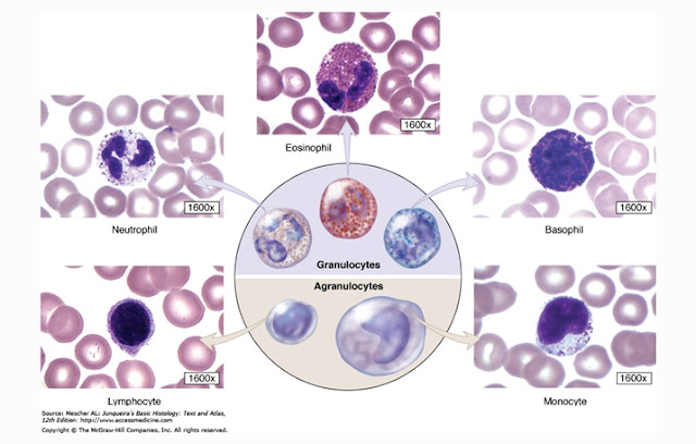
White blood cells according to the shape on their surface is divided into two groups, polymorphonuclear cells (granulated) and mononuclear cells (not granulated). Both groups are divided into several forms,:
1. polymorphonuclear cells
  • Basophils
  • Eosinophils
  • Neutrophils

2. Mononuclear Cells
  • Lymphocytes
  • Monocytes
Definition, Structure and Function of Leukocytes (White Blood Cells)
LEUKOCYTES (WHITE BLOOD CELLS)
Here is a detailed explanation of the types of red blood cells which have been mentioned above,:
1. Basophils
Basophils are types of white blood cells that have the percentage 0.01-0.03% of the total number of white blood cell types. As mentioned above, basophils have granules in their cytoplasm. The white blood cells of this type has two lobes. Basophil itself is one of a group of granulocytes to emerge towards certain body tissues.

Basophil works when there is event of an allergic reaction in our body. So with these allergic reactions, basophil going out and capture the allergen and secrete histamine. Histamine will cause the blood vessels dilate (bigger). The more allergen that enters the body, the more number of basophil working against him. The characteristics of basophil cells are:
  • Has phagocytes Properties
  • Between 12-15 micrometers in diameter
  • Having a number of 0.01-0.03% per millimeter of blood
  • Having a large granule
  • Formed in the bone marrow
  • U-shaped and speckled
  • Blue in color
  • Had a core that is not segmented

2. Eosinophils
Eosinophils are a type of white blood cell that has the amount of 7% of the total white blood cell. These cells have a  diameter of 10-12 micrometers. These cells will be increased in the event of reaction associated with asthma, allergies, and fever.
Eosinophils have a duty to combat the reactions from the allergen within 8-12 days. Eosinophils have chemical substances, such as ribonuclease, histamine lipase, and others. The characteristics of eosinophil are:
  • Are phagocytes and also acidic
  • Has a core with two lobes
  • Red in color
  • The core is in the middle of the cell
  • Having a rough and dense granules
  • Having a life span of between 8-12 days
  • It looked like a ball and its size is about 9 mm
  • Formed in the bone marrow

3. Neutrophils
Neutrophil white blood cell types are cells that have the percentage of 50-60% in the blood. Neutrophils have three nucleus. These cells act as a first barrier (defense) in response to allergic reactions in the body. Neutrophil is a phagocytic cells so can they fight the problem using various chemicals such as hydrogen peroxide, free radicals, and hypochlorite. Neutrophils also play a role in the inflammatory process, and will increase if the more inflammatory agent are involved. The characteristics possessed by neutrophil cells are:
  • Having 3 nucleus
  • Has a size of approximately 8 mm
  • Having a life time between 6-20 hours
  • Properties phagocytes
  • Neutrophil production output amounted to 100 billion cells per day

4. Lymphocytes
Types of white blood cells called lymphocytes has overall amount of 20-25% of the total of all types of white blood cells in the blood. The body produces lymphocytes in the bone marrow and spleen. This type is divided into large and small lymphocytes. The difference between the two is, small lymphocytes have a darker core that almost filled the entire cytoplasm, whereas in large lymphocytes, essentially pale and have a smaller size and does not fill all the cytoplasm.

There are approximately 8000 lmphocytes cells in the white blood cells. If an increase in the levels of lymphocyte count, it indicates the occurrence of a process called leukemia. There are 5 types of lymphocytes, the B lymphocytes, T helper cells, Cytotoxic T cells, memory T cells, and suppressor T cells. The characteristics of lymphocytes are:
  • Not having granules in the cytoplasm and size vary from small to large
  • Dark colored core surrounded by a narrow cytoplasmic ring
  • Age range from days to several months
  • If the body is exposed by pathogen, the B lymphocyte cells will form plasma cells in the tissues

5. Monocytes
Monocytes are the largest leukocytes that dont have granules. Their shape varies, round or oval ,grooved or shape of a horseshoe. monocytes Color are paler than the lymphocytes core. Monocytes form the approximately 3-8% of leukocytes in the blood.
The mechanism to fight the foreign objects made by monocytes is to eat it, despite its larger size. Monocytes will turn into macrophages inside certain tissue for a mechanism of resistance. The characteristics of monocytes are:
  • The agranular leukocytes characterized by a horseshoe-shaped nucleus
  • Life in the connective tissue for months
  • There are 3-8% monocytes of leukocytes in the blood
  • Have a peanuts-like shape

D. HOW DOES LEUKOCYTES WORKS (WHITE BLOOD CELL)
At the time of foreign substances or microorganisms that can harm the body entering our body, chemical signals will tell monocytes, so the monocytes will immediately move out of the blood vessels to where the attack occurred. Monocytes will fight foreign substances so that monocytes secrete substances called cylocsin awaken the body's fever.

Monocytes can not work alone, but monocytes assisted by neutrophils. Resistance conducted by neutrophils, monocytes and neutrophils often cause death of neutrophils. However, neutrophils sacrifice will not be in vain, because when they die, it will form pus as a mechanism for inhibiting the onset of subsequent infection.

After that, the monocytes had killed a foreign substance will carries this foreign substance into lymphocytes. Lymphocyte function to record what kinds of germs are killed by monocytes and then establish a defense system (antibodies) against the substance, so that when the substances back into the body in the future, it would be easy to resist.

Lymphocytes has two different types of cells that responsible for this job. The first is as a specialist agency B cells that release antibodies to detect the presence of bacteria that have been recorded, and will be fighting in the distance. While the T cells would maintain or oversee the antibody and keep them at the surface of the cell.

E. THE DAMAGE LEUKOCYTES (WHITE BLOOD CELLS)
Levels of white blood cells in the human body is different, but in general, there are normal levels of white blood cells present in the body of a healthy person, that is 4000 per microliter of blood. Circumstances that lead to low white blood cells is called leukopenia, while high white blood cells is called leukocytosis.
Causes that can cause of the decreasing levels of white blood cells in the body of a healthy person are:
  • A viral infection that affects the bone marrow
  • Congenital disorder that affects the bone marrow
  • Cancer that affects the bone marrow
  • Drugs that work by suppressing the levels of white blood cells in the blood
  • Severe infection that cause the death of white blood cells faster than usual.
  • Diseases that cause low white blood cells such as HIV / AIDS, chemotherapy, leukemia, lupus, myelodysplasia syndromes, aplastic anemia, certain drugs such as antibiotics, diuretics, hiperslenisme, Kostmann syndrome, radiation therapy, and vitamin deficiencies.

While high levels of white blood cells circulating in the blood may be caused by:
  • Infections that can increase the production of white blood cells
  • Abnormal function of the bone marrow resulting in higher production of white blood cells
  • Reactions to certain drugs
  • Some disease that can cause high white blood cell in the blood such as leukemia limpotik acute or chronic, acute myeloid leukemia or chronic, drugs such as epinephrine, bacterial or viral infections, Myelofibrosis, a severe allergic reaction, stress, tuberculosis, whooping cough, and smoking.

Medulla Oblongata : Definition, Structure and Functions

A. UNDERSTANDING THE DEFINITION OF MEDULLA OBLONGATA
The brain is divided into five sections, namely a Cerebrum and cerebellum, midbrain, pons and medulla oblongata. Under the cerebellum are pons which serves as the respiratory center. Well, under the pons, there is an organ called the medulla oblongata.

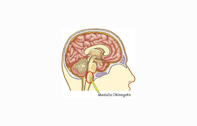
Medulla oblongata is one part of the brainstem that are Located under the pons. Medulla oblongata itself plays a role in controlling the functions of the autonomic (involuntary functions) system such as breathing, digestion, heart rate, vascular function, as well as swallowing and sneezing.
Medulla oblongata is also an organ that can deliver signals from the brain before it is submitted to the nerves in the spine (spinal cord). Therefore, the medulla oblongata is a very important organ in the human body.
Definition, Structure and Functions of Medulla Oblongata
MEDULLA OBLONGATA
B. THE STRUCTURE OF MEDULLA OBLONGATA
Medulla oblongata (Cord marrow / cord connection) has some structure / part which has the characteristics and duties of each, these sections are:
1. Descending Tract
Descending tracts are neural pathways from the brain and pass through to the medulla oblongata and the to the spinal cord. Descending tract functions is to carry impulses (signals) received from brain nerve to be delivered to the spinal cord before continuing to the organs of the body.

2. Ascending Tract
Ascending tract is the opposite of the descending tract. If the descending tract comes from the brain, the ascending tracts coming from the spinal cord, and serves to carry impulses (signals) from the organs through the spinal cord, and pass through the medulla oblongata then to the brain.

3. Spinal Nerves Nucleus
There are three nucleus (core) nerve in the medulla oblongata, the hipoglossus nucleus (which is the core of the nerve to innervate part of the tongue), motoric dorsal nucleus of the vagus (to control the motoric movement) and the nucleus of the solitaries tract.
Parts of Medulla Oblongata
PARTS OF MEDULLA OBLONGATA
C. THE FUNCTIONS OF MEDULLA OBLONGATA
As mentioned before, the medulla oblongata plays a role in controlling the body's autonomic functions such as breathing rhythm control, set the rhythm of digestion, regulate heart rhythm, regulates the function of blood vessels, and others. These functions are influenced by several unique receptors located throughout the body and will react to environmental changes. For example, the chemoreceptors in the lungs that will send information to the medulla oblongata when the body is exercising, the medulla oblongata will send a chemoreceptors that improve the body's oxygen uptake in order to survive in this situation. These kind of receptors will control medulla oblongata in order to regulate the respiratory rhythm in accordance with the condition of the body.

There are a lot of medulla oblongata’s function that we still dont know. But for sure, here are some of the functions of the medulla oblongata :
  • As a connection between the brain and spinal cord
  • Regulate the body's reflex
  • Stimulate thirst
  • regulate emotions
  • Responsible for some of autonomic function
  • Affect hormone production in the pituitary gland in the brain
  • Controlling bed activities
  • Controlling the diameter of blood vessels, widened or narrowed in accordance with the state of the body
  • Detect the degree of acidity of the blood
  • As a central regulator of heart rate
  • Assist breathing rhythm
  • Regulate blood circulation
  • Regulates body temperature

How Does medulla oblongata works ?
1. Parts of Medulla oblongata that serves as the respiratory center:
Dorsal Group, the nervous system that forms the automatic breathing.
Ventral Group, a group of nervous system which innervate the muscles of respiration

2. The medulla oblongata as regulator of the Heart
Cardioaccelerator Center, which works to increase heart rate and strength of heart contractions
Cardioinhibitori Center, which works to reduce heart beat to the vagus nerve (parasympathetic nerve)

3. The medulla oblongata as Vasomotor Center
Medulla as vasomotor means that medulla serves to control the diameter of our blood vessel function through sympathetic nerves in the measurement of blood pressure

4. The medulla oblongata as Non-Vital Reflex Center
In this case, the medulla oblongata function is to regulation of swallowing, vomiting, coughing, sneezing, choked.

D. MEDULLA OBLONGATA CIRCULATION SYSTEM
Medulla oblongata is just like other organs in the human body, it has a special blood vessels that carry blood to the medulla oblongata itself. There are several branches of the blood vessels that circulate medulla oblongata, which are:
1. Anterior Spinal Artery
These arteries supplying blood and nutrition for almost all part of meudulla oblongata.
2. Posterior Inferior Cerebellar Artery (PICA)
This artery is a major branch associated with anterior spinal artery. These blood vessels provide blood supply to the back side (posterolateral) of medulla oblongata.

Tendon : Definition, Structure and Functions

A. UNDERSTANDING THE DEFINITION OF TENDON
Tendons are connective tissues that connect bone to muscle. Tendon serves to balance the bone and muscle when the movement process. Tendon that attached to the bone and mobile called insersio (insertian = inset), while the tendon attached to the bone but immobile called Origo (origin = origin). The structure of tendon at the tip muscles is small, tough and hard.
Definition, Structure and Functions of Tendon
TENDON
B. THE FUNCTIONS OF TENDON
As we have explained previously, the main function of the tendon is to control the movement mechanism (effective and efficient). When the movement occur, the tendon will adapt to the changing position of the bones and muscles so that the movement is perfect. Tendon function is associated with the contraction (shortening) and relaxation (elongation) of muscle. When the muscles are contract, the tendons transmit energy from the contraction to the bones and joints, while the muscles are relax, tendons will also adjust its state. Although it has a very strong structure, tendon can also be damaged. Most often Injuries to the tendon due to excessive training or accident.

C. THE STRUCTURE OF TENDON
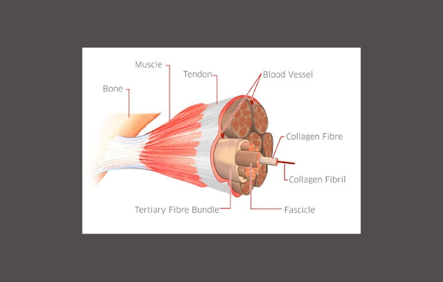
Please note the above image to make it easier to understand our explanation
We will try to explain the structure of the tendon form the smallest constituent components.
The smallest structure of tendon is collagen fibrils. These fibrils are solid, strong and flexible. The nature of the collagen fibers would make it resistant to traction and encouragement between the bones and muscles. The basic constituent molecules of collagen fibrils is from some tropokolagen united to form microfibrils and microfibrils of collagen are combined together to form subfibril collagen, and then combined to form collagen fibrils.
Structure of Tendon
TENDON STRUCTURE
The combination of collagen fibrils will merge and form a protective layer in the form of strands called collagen fibers.
Then the combination of collagen fibers that are coated by a endotendon layer will form Primary Collagen Fiber (Sub-fasicle).
The primary will be joining to each other and coated by endotendon layer (layer that serves to protect and stabilize the tendon) to form Secondary collagen fiber bundles (fasicle).
A combination of several secondary collagen fibers that coated by endotendon layer (layer that serves to protect and stabilize the tendon) formed Tertiary Collagen Fiber Bundles.
Now a collection of Tertiary Collagen Fiber Bundles that coated by epitendon layer (the outer layer of the tendon) will form the complete Tendon. 

Liver (Hepar) : Definition, Structure and Functions

A. UNDERSTANDING THE DEFINITION OF LIVER (HEPAR)
Liver (Hepar) is one of the largest glandular organ in the human body. The liver is located in the upper right abdomen . The color is dark red-brown and is the organ that has many functions in supporting life. Liver’s weight is about 3-5% of a person's body weight.
Definition, Structure, and Functions of Liver (Hepar)
LIVER (HEPAR)
B. THE FUNCTIONS OF LIVER (HEPAR)
  • Vascular function, ie for storage and filtration of blood.
  • Secretion function, to produce urea and bile salts were later distributed through channels (ducts) into the digestive tract.
  • Metabolic function, liver also has the function of the metabolism of carbohydrates, protein, fat, and other body parts.
  • Storage functions, the liver can store fat, glycogen, vitamins, and iron. Then will be issued when needed by the body.
  • Antidote function, when we consume food or drinks that contain toxins, the liver will transform the toxic components, so that components can be used or removed from the body. For example, the results of carbohydrate metabolism produces lactic acid is converted into glycogen so that it can be used for energy reserves. Another example is the metabolism of proteins that produce ammonia is converted into urea and excreted from the body in the urine through the process of urination.

C. STRUCTURE AND PARTS OF THE LIVER (HEPAR)
The liver consists of two main parts, parts of the liver called right and left lobe. Right and left lobes are separated by a fissure which is formed from a combination of several ligaments. Each lobe is composed by functional units called hexagonal lobules. At the center of the lobules there are vessels of the vein or the central vein. Each central vein will then be joined together to form the hepatic vein blood vessel which is the main veins of the liver. The structure that separates each the lobules with other lobules is called lacunae. The tissue is composed by lobolus cells called hepatocytes. Each cells separated by hepatocytes canaliculi, then every canaliculi will merge to form bile ducts.
Liver Blood Supply
LIVER BLOOD SUPPLY
Supply of nutrients and oxygen to the liver (hepar) is obtained from the heart, same as other organs. However, blood flow of hepar is a bit different with other organs. The liver receives blood from two main sources, namely the hepatic artery and hepatic portal vein.
Hepatic Arteries carry oxygen-rich blood and nutrients from the heart and supply about 20% of the hepatic blood.
While the hepatic portal vein carries blood from the digestive system. This blood contains the products of metabolism and digestion. Portal vein blood supplies about 80% of the liver.
Then the blood will come out of the central vein to the hepatic vein and inferior vena cava.
To understand this explanation please see the picture below:

BLOOD VESSELS OF LIVER (HEPAR)

Tooth : Definition, Structure, Functions, Types

A. UNDERSTANDING THE DEFINITION OF TOOTH
Tooth is the organ of the digestive system located in the oral cavity. Tooth functioning as mechanical digestion tools because it serves to cut, rip, and chew food before the food gets into the next digestive organ. Teeth have dense and hard structure to perform its functions properly.
Definition, Structure and Functions of Tooth
TOOTH
B. PARTS TEETH
Teeth are composed of three main parts,:
1. Dental Crown (Peak)
Dental crown is a part that is visible from outside. This layer looks white because it is coated by a structure called the enamel. Each type of tooth has a different shape of crown.

2. Dental Neck (Colum)
Neck or colum is the part that is built into the gums. This section is a barrier between the crown and the root.

3. Tooth Root
Tooth root is the part that is embedded in the jaw (see picture) so it is not visible from the outside. Humans have some types of teeth, every tooth can have a number of different roots, some have two roots and some other have three roots.
Structure and Parts of The Tooth
TOOTH STRUCTURE
C. THE STRUCTURE OF TOOTH
These structure is layers that forms teeth :
1. Enamel
As we have explained before, the enamel is a layer that coat the tooth crown. The enamel had harsh nature because it is formed by calcium. At the top part the enamel layer is the hardest part, the bottom part will be thin so this part is not too hard. Actually, the color of the enamel is not fully white, but grayish. The function of this layer is to protect the teeth bone from the outside.

2. Dental cementum
The root of the tooth in the jaw lined by this cementum. This layer also serves to protect the roots of the teeth, but the structure is not as thick and hard as enamel. Under normal circumstances this layer is not visible from the outside because it is covered by the jaw and gums, but some diseases can cause visible cementum.

3. Dental Bone
This layer is located after the enamel on the crown and after the cement layer on the root of the tooth. Dental bone is the widest part of the tooth, its structure covering the entire length of the tooth, from crown to root. It is named “Dental Bone” because its structure is similar to the bone but harder because it has more calcium content. Bone also serves as a protection, second after enamel and tooth cementum. Colors of this section are usually yellowish.

4. Dental Cavities (pulp)
The pulp is the soft tissue that forms on the inside of the tooth cavity. This cavity contains blood vessels and nerve vessels. The pulp serves as a conduit for dental nutrition because they have no blood vessels, and to identify foreign objects, pressure, temperature and pain because they have nerve vessels.

D. TYPES OF THE TEETH
1. Based on the shape and function, human teeth divided into four kinds :
a. Incisors
Incisors are the teeth that serve to cut off food. Incisor crown-shaped “upright” with the tip of his teeth shaped like a shovel. Incisors located on the front of the tongue and have only one root. Adult human has 4 incisors, 2 in the lower jaw, and 2 in the upper jaw.

b. Canine
Canine teeth are teeth that serve to tear and rip the food. Canines is sharp. Canines located after the incisors. Adult human have 4 pieces canines, 2 situated in the lower jaw and the other two located in the upper jaw.

c. Premolar
Premolars front teeth used to grind and chew food. Premolar tooth-shaped upright somewhat wider and lower than the two types before. Premolar tooth had a few bumps in the crown. Front molars have two pieces of root. In adult humans there are 8 pieces of the premolar teeth 4 the lower jaw and 4 in the upper jaw.

d. Molar
Molars serve to crush, pulverize, crush and grind food. Molar teeth shaped like premolar teeth but have a few bumps on the crown. Most molars have three roots, but there are also some which only has two roots. Adult human has 12 pieces molars teeth, ie 6 pieces in the upper jaw and the other six in the lower jaw.
Incisor, Canine, Premolar, Molar
TYPES OF TOOTH
2. Types of Dental by Age teeth were divided into two, namely:
a. Deciduous teeth (Temporary Teeth/Milk Teeth)
Deciduous teeth are teeth that start growing in humans at about the age of 6 months. Teeth are generally growing incisors first is located at the center of the jaw. As humans age, one by one of these deciduous teeth are replaced by permanent teeth. Generally, teeth start date at children aged 4-6 years. There are 20 pieces Deciduous teeth, 8 pieces incisors, four canines and eight premolars.

b. Permanent teeth
Permanent teeth is teeth that replace the milk teeth and will be the teeth for a lifetime. Permanent teeth will not erupt, unless there is an abnormality. Overall number of permanent teeth is 32 pieces, : eight incisors, four canines, eight premolars and 12 molars.

E. STRUCTURE AROUND THE TEETH
  • Periodontal ligament, the connective tissue structure that is attached to the roots of the teeth and is attached to the bone. The periodontal ligament will continue as the gingival tissue (gums) and connected with blood vessels in the bone.
  • Oral mucosa, is oral cavity that is coated by stratified squamous epithelium cells. Oral mucosal main function is to protect tissue or other organs in the oral cavity.
  • Gingiva (gums), an oral mucosa covering the maxillary bone and mandibula in the oral cavity. Gingival tissue itself is actually transparent in color, the red color is formed because there are many blood vessels in that section. The function is to protect the gums and tissues surrounding the tooth root.
  • Nerves and blood vessels, around the teeth also are nerves and blood vessels. Neural tube serves to receive and provide a stimulus, whereas blood vessels serves to provide the intake of nutrients needed by the surrounding structures.